Quick Links
Breast Cancer Links
- Breast Cancer Overview
- Breast Cancer Resources
- IHC Atlas
- Clinical Case Reviews
- Spotlight Series
- Knowledge Check
Gastric Cancer Links

Welcome to the new, updated HER2Know
We have expanded our resource library so you can now explore content on HER2 in tumors beyond breast.
Looking for the original HER2Know breast cancer resources?
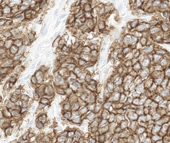
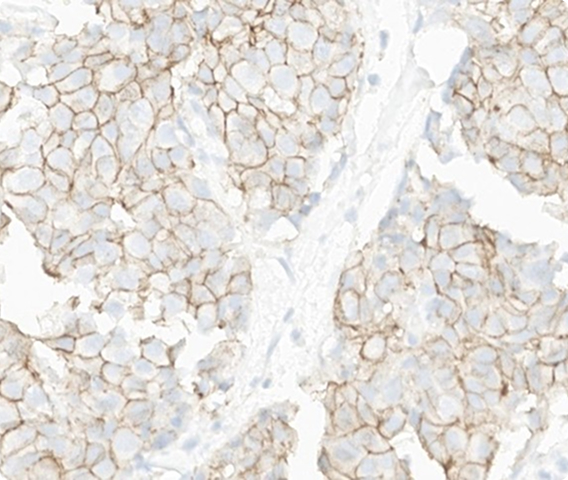
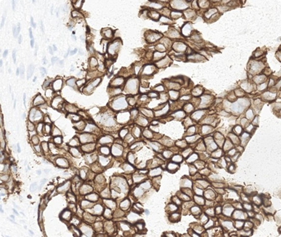
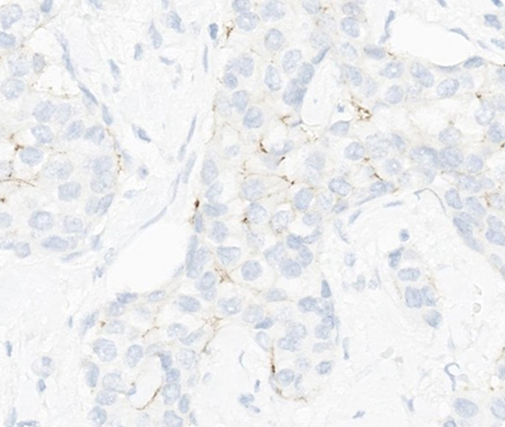
Assess HER2 IHC clinical cases and see how your scores compare to your peers.
Put your skills to the test as you examine and classify clinical breast cancer cases curated and consensus-scored by our Pathology Faculty. Each question features whole slide images and interpretation summaries, helping you take your practice to the next level.
Which HER2 IHC score would you assign to this slide?
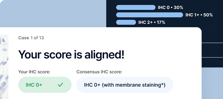
View whole slide imageYour score is aligned!
Your IHC score:
IHC 0+
Consensus IHC score:
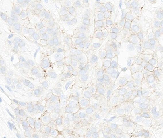
IHC 0+ (with membrane staining*)
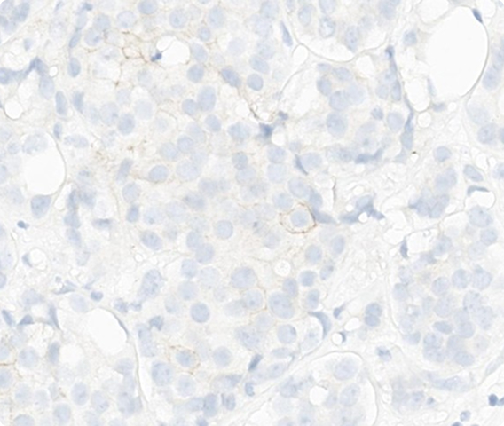
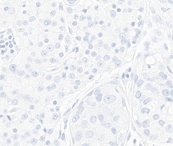
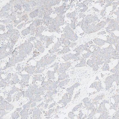
This case shows membrane staining is incomplete and faint/barely perceptible in ≤10% of invasive tumor cells.
*According to the CAP Breast Biomarker Reporting Template, a score of IHC 0+ has membrane staining that is incomplete and is faint/barely perceptible and in ≤10% of tumor cells.1
View whole slide imageNot aligned with the Pathology Faculty score
Your IHC score:
IHC 0
Consensus IHC score:
IHC 0 (no membrane staining)
This case shows membrane staining is incomplete and faint/barely perceptible in ≤10% of invasive tumor cells.
*According to the CAP Breast Biomarker Reporting Template, a score of IHC 0+ has membrane staining that is incomplete and is faint/barely perceptible and in ≤10% of tumor cells.1
View whole slide image
Which HER2 IHC score would you assign to this slide?
View whole slide imageYour score is aligned!
Your IHC score:
IHC 2+
Consensus IHC score:
IHC 2+
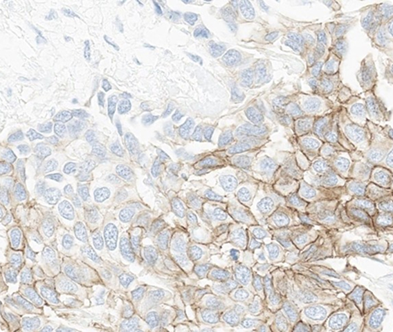
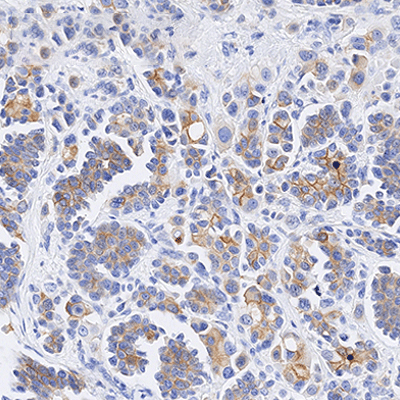
This case shows weak to moderate complete membrane staining in >10% of invasive tumor cells.
Reflex ISH testing is required to determine HER2 amplification status.
Not aligned with the Pathology Faculty score
Your IHC score:
IHC 2+
Consensus IHC score:
IHC 2+
This case shows weak to moderate complete membrane staining in >10% of invasive tumor cells.
Reflex ISH testing is required to determine HER2 amplification status. If ISH is negative, this case falls under “HER2 low” category per DESTINY-Breast trials and may be eligible for fam-trastuzumab deruxtecan-nxki in the metastatic setting.
Which HER2 IHC score would you assign to this slide?
View whole slide imageYour score is aligned!
Your IHC score:
IHC 1+
Consensus IHC score:
IHC 1+
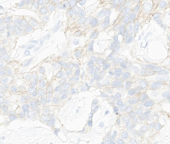
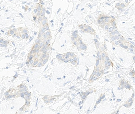
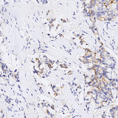
This case shows incomplete membrane staining that is faint/barely perceptible in >10% of invasive tumor cells, with focal weak to moderate complete membranous staining in ≤10% of tumor cells.
View whole slide imageNot aligned with the Pathology Faculty score
Your IHC score:
IHC 1+
Consensus IHC score:
IHC 1+
This case shows incomplete membrane staining that is faint/barely perceptible in >10% of invasive tumor cells, with focal weak to moderate complete membranous staining in ≤10% of tumor cells.
View whole slide image
Which HER2 IHC score would you assign to this slide?
View whole slide imageYour score is aligned!
Your IHC score:
IHC 0
Consensus IHC score:
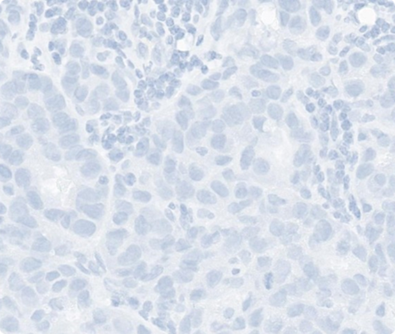
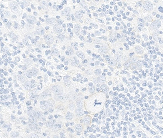
IHC 0 (no membrane staining)
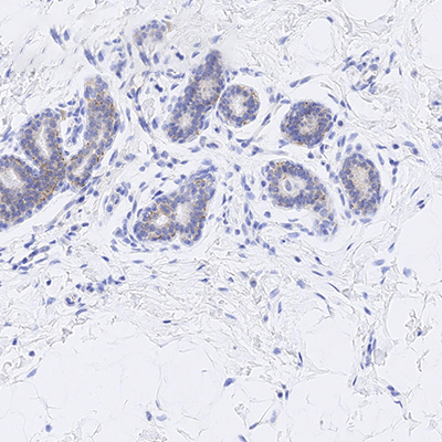
This case shows no membrane staining detected in invasive tumor cells. Internal control (benign lobule) shows expected faint membranous staining, confirming assay performance.
This slide shows absent membranous staining throughout the invasive tumor cells. In evaluating such cases, it is critical to assess both internal and on-slide positive controls. In this case, a benign lobule with faint membranous staining on the left side of the specimen serves as an internal control. This case also requires careful distinction between invasive and in situ carcinoma.
Not aligned with the Pathology Faculty score
Your IHC score:
IHC 0
Consensus IHC score:
IHC 0 (no membrane staining)
This case shows no membrane staining detected in invasive tumor cells. Internal control (benign lobule) shows expected faint membranous staining, confirming assay performance.
This slide shows absent membranous staining throughout the invasive tumor cells. In evaluating such cases, it is critical to assess both internal and on-slide positive controls. In this case, a benign lobule with faint membranous staining on the left side of the specimen serves as an internal control. This case also requires careful distinction between invasive and in situ carcinoma.
Which HER2 IHC score would you assign to this slide?
View whole slide imageYour score is aligned!
Your IHC score:
IHC 0
Consensus IHC score:
IHC 0 (no membrane staining)
This case shows no membrane staining detected in invasive tumor cells.
This case highlights the importance of distinguishing invasive from in situ carcinoma, as only invasive tumor cells should be evaluated for HER2 membranous staining.
Not aligned with the Pathology Faculty score
Your IHC score:
IHC 0
Consensus IHC score:
IHC 0 (no membrane staining)
This case shows no membrane staining detected in invasive tumor cells.
This case highlights the importance of distinguishing invasive from in situ carcinoma, as only invasive tumor cells should be evaluated for HER2 membranous staining.
Which HER2 IHC score would you assign to this slide?
View whole slide imageYour score is aligned!
Your IHC score:
IHC 3+
Consensus IHC score:
IHC 3+
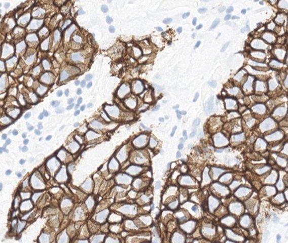
This case shows circumferential membrane staining that is complete, intense, and in >10% of invasive tumor cells.
View whole slide imageNot aligned with the Pathology Faculty score
Your IHC score:
IHC 3+
Consensus IHC score:
IHC 3+
This case shows circumferential membrane staining that is complete, intense, and in >10% of invasive tumor cells.
View whole slide image
Which HER2 IHC score would you assign to this slide?
View whole slide imageYour score is aligned!
Your IHC score:
IHC 1+
Consensus IHC score:
IHC 1+
This case shows incomplete membrane staining that is faint/barely perceptible in >10% of invasive tumor cells.
The invasive component involves a small portion of the tissue. Remaining tissue includes ductal carcinoma in situ and benign glandular elements. A duct with apocrine metaplasia shows granular cytoplasmic staining, which should not be interpreted as HER2 positivity. Careful distinction between invasive carcinoma and non-invasive elements is essential.
Not aligned with the Pathology Faculty score
Your IHC score:
IHC 1+
Consensus IHC score:
IHC 1+
This case shows incomplete membrane staining that is faint/barely perceptible in >10% of invasive tumor cells.
The invasive component involves a small portion of the tissue. Remaining tissue includes ductal carcinoma in situ and benign glandular elements. A duct with apocrine metaplasia shows granular cytoplasmic staining, which should not be interpreted as HER2 positivity. Careful distinction between invasive carcinoma and non-invasive elements is essential.
Which HER2 IHC score would you assign to this slide?
View whole slide imageYour score is aligned!
Your IHC score:
IHC 3+
Consensus IHC score:
IHC 3+
This case shows circumferential membrane staining that is complete, intense, and in >10% of invasive tumor cells.
View whole slide imageNot aligned with the Pathology Faculty score
Your IHC score:
IHC 3+
Consensus IHC score:
IHC 3+
This case shows circumferential membrane staining that is complete, intense, and in >10% of invasive tumor cells.
View whole slide image
Which HER2 IHC score would you assign to this slide?
View whole slide imageYour score is aligned!
Your IHC score:
IHC 2+
Consensus IHC score:
IHC 2+
This case shows weak to moderate complete membrane staining in >10% of invasive tumor cells, with areas of incomplete weak staining.
Reflex ISH testing is required to determine HER2 amplification status.
Not aligned with the Pathology Faculty score
Your IHC score:
IHC 2+
Consensus IHC score:
IHC 2+
This case shows weak to moderate complete membrane staining in >10% of invasive tumor cells, with areas of incomplete weak staining.
Reflex ISH testing is required to determine HER2 amplification status. If ISH is negative, this case falls under “HER2 low” category per DESTINY-Breast trials and may be eligible for fam-trastuzumab deruxtecan-nxki in the metastatic setting.
Which HER2 IHC score would you assign to this slide?
View whole slide imageYour score is aligned!
Your IHC score:
IHC 3+
Consensus IHC score:
IHC 3+
This case demonstrates strong, complete circumferential staining in the invasive tumor cells.
View whole slide imageNot aligned with the Pathology Faculty score
Your IHC score:
IHC 3+
Consensus IHC score:
IHC 3+
This case demonstrates strong, complete circumferential staining in the invasive tumor cells.
View whole slide image
Which HER2 IHC score would you assign to this slide?
View whole slide imageYour score is aligned!
Your IHC score:
IHC 0+
Consensus IHC score:
IHC 0+ (with membrane staining*)
This case shows membrane staining that is incomplete and faint/barely perceptible in ≤10% of invasive tumor cells.
*According to the CAP Breast Biomarker Reporting Template, a score of IHC 0+ has membrane staining that is incomplete and is faint/barely perceptible and in ≤10% of tumor cells.1
View whole slide imageNot aligned with the Pathology Faculty score
Your IHC score:
IHC 0+
Consensus IHC score:
IHC 0+ (with membrane staining*)
This case shows membrane staining that is incomplete and faint/barely perceptible in ≤10% of invasive tumor cells.
*According to the CAP Breast Biomarker Reporting Template, a score of IHC 0+ has membrane staining that is incomplete and is faint/barely perceptible and in ≤10% of tumor cells.1
View whole slide image
Which HER2 IHC score would you assign to this slide?
View whole slide imageYour score is aligned!
Your IHC score:
IHC 1+
Consensus IHC score:
IHC 1+
This case shows incomplete, faint/barely perceptible membrane staining in >10% of invasive tumor cells. This case shows predominantly weak membranous staining in the invasive tumor cells.
Caution should be taken not to interpret luminal and/or granular cytoplasmic staining as positive membranous staining.
Not aligned with the Pathology Faculty score
Your IHC score:
IHC 1+
Consensus IHC score:
IHC 1+
This case shows incomplete, faint/barely perceptible membrane staining in >10% of invasive tumor cells. This case shows predominantly weak membranous staining in the invasive tumor cells.
Caution should be taken not to interpret luminal and/or granular cytoplasmic staining as positive membranous staining.
Which HER2 IHC score would you assign to this slide?
View whole slide imageYour score is aligned!
Your IHC score:
IHC 0+
Consensus IHC score:
IHC 0+ (with membrane staining*)
This case shows membrane staining that is incomplete and faint/barely perceptible in ≤10% of invasive tumor cells.
*According to the CAP Breast Biomarker Reporting Template, a score of IHC 0+ has membrane staining that is incomplete and is faint/barely perceptible and in ≤10% of tumor cells.1
View whole slide imageNot aligned with the Pathology Faculty score
Your IHC score:
IHC 0+
Consensus IHC score:
IHC 0+ (with membrane staining*)
This case shows membrane staining that is incomplete and faint/barely perceptible in ≤10% of invasive tumor cells (pattern consistent with HER2 ultralow).
*According to the CAP Breast Biomarker Reporting Template, a score of IHC 0+ has membrane staining that is incomplete and is faint/barely perceptible and in ≤10% of tumor cells.1
View whole slide imageHow does your HER2 IHC scoring compare to your fellow pathologists? Examine these whole slide images and select your score.
Slide 1 OF 13
Which HER2 IHC score would you assign to this case?
Service down. Please try again some time
How of your peers scored this slide
Slide 2 OF 13
Which HER2 IHC score would you assign to this case?
Service down. Please try again some time
How of your peers scored this slide
Slide 3 OF 13
Which HER2 IHC score would you assign to this case?
Service down. Please try again some time
How of your peers scored this slide
Slide 4 OF 13
Which HER2 IHC score would you assign to this case?
Service down. Please try again some time
How of your peers scored this slide
Slide 5 OF 13
Which HER2 IHC score would you assign to this case?
Service down. Please try again some time
How of your peers scored this slide
Slide 6 OF 13
Which HER2 IHC score would you assign to this case?
Service down. Please try again some time
How of your peers scored this slide
Slide 7 OF 13
Which HER2 IHC score would you assign to this case?
Service down. Please try again some time
How of your peers scored this slide
Slide 8 OF 13
Which HER2 IHC score would you assign to this case?
Service down. Please try again some time
How of your peers scored this slide
Slide 9 OF 13
Which HER2 IHC score would you assign to this case?
Service down. Please try again some time
How of your peers scored this slide
Slide 10 OF 13
Which HER2 IHC score would you assign to this case?
Service down. Please try again some time
How of your peers scored this slide
Slide 11 OF 13
Which HER2 IHC score would you assign to this case?
Service down. Please try again some time
How of your peers scored this slide
Slide 12 OF 13
Which HER2 IHC score would you assign to this case?
Service down. Please try again some time
How of your peers scored this slide
Slide 13 OF 13
Which HER2 IHC score would you assign to this case?
Service down. Please try again some time
How of your peers scored this slide
ASCO, American Society of Clinical Oncology; CAP, College of American Pathologists; HER2, human epidermal growth factor receptor 2; IHC, immunohistochemistry.
1. College of American Pathologists. Template for reporting results of biomarker testing of specimens from patients with carcinoma of the breast. Accessed June 12, 2025. https://www.cap.org/wp-content/uploads/2026/07/Breast.Bmk_1.6.1.0.-REL_CAPCP-1.pdf
©2026 Daiichi Sankyo, Inc. and AstraZeneca. All rights reserved.
PP-US-8201a-2774-1, US-109020, PP-US-8201a-1849, US-84238, PP-US-8201a-2323, US-99813 07/26
Request an AstraZeneca Medical Affairs Representative Follow-Up