



Quick Links
Breast Cancer Links
- Breast Cancer Overview
- Breast Cancer Resources
- IHC Atlas
- Clinical Case Reviews
- Spotlight Series
- Knowledge Check


Welcome to the new, updated HER2Know
We have expanded our .
Looking for the original HER2Know breast cancer resources?


Shi Wei, MD, PhD, is the Professor with Tenure and the Barbara F. Atkinson Endowed Professor, Director of Translational Research at the Department of Pathology and Laboratory Medicine, University of Kansas Medical Center. In this article, Dr. Wei, MD, PhD, discusses the use of bone specimens as samples for HER2 IHC in metastatic breast cancer. He considers the impact of the decalcification procedure on the reliability of test results, establishing the advantages and disadvantages of the different procedures and reagents.
It is quite common to receive bone biopsy specimens from patients with a known history of breast cancer for confirmation of metastatic disease, as well as biomarker studies for systemic treatment decision making.1 Moreover, it is not uncommon to encounter a bone biopsy from patients without a prior history of malignancy, in whom metastatic disease is the initial presentation of breast cancer.1 Therefore, IHC testing for HER2 on bone specimens is frequently performed in routine pathology practice.
Decalcification is a technique for removing minerals from bone or other calcified tissue to obtain high-quality paraffin sections.2 It is carried out after thorough fixation and prior to processing in paraffin.2
“It is important to validate the decalcification methods to ensure that they do not result in clinically relevant discordance.”
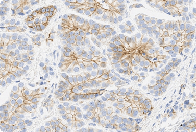
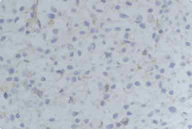
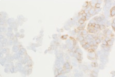
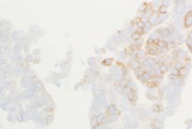
There are different types of decalcifying agents including strong mineral acids, weaker organic acids, and chelating agents.3 Decalcification using strong acids (such as hydrochloric acid) is not recommended, as it can lead to rapid tissue degradation and loss of nuclear staining, as such their use should be limited.3 Weak acids (such as formic acid) are less likely to interfere with immunostaining as they are slower acting and therefore are used more widely for decalcification.3 Decalcification using chelating agents (such as EDTA) best preserves histomorphology and generally can be more reliably used for IHC, FISH, or PCR (Figure 1).3,4 However, decalcification using chelating agents is more time-consuming and therefore may not be appropriate for processing urgent specimens.3 The decalcification method of bone specimens is not standardized across the pathology community, therefore it is of great importance to validate decalcifications internally.2
“It is recognized that hydrochloric acid-based and long-term formic acid-based decalcification may induce false-negative results on IHC.”2,3
In one analysis it was reported that when compared to acetic and hydrochloric/formic acid, EDTA decalcification minimally affects ER, PR, and HER2 results. Although, in this particular case the comparison was between the primary tumor and bone metastasis, not the same tissue with and without decalcification.2 In contrast, another study demonstrated that mean IHC scores for several nuclear and membranous/cytoplasmic antigens (including HER2) were similar with formic acid, EDTA, and non-decalcified samples.3 Note that the duration of decalcification was different for various decalcifying agents and across each of the studies.3
Within CAP reporting templates for biomarker reporting there is additional guidance wherein cases where pre-analytical variables may have an impact on test results, the variables must be reported. This includes decalcification of bone specimens as this could potentially alter immunoreactivity.6 In all cases, if tests undergo technical issues that prevent results being reported as positive, negative, or equivocal, results should be reported as indeterminate, and another sample should be requested for testing. This includes cases with inadequate specimen handling, where artifacts make interpretation difficult, or analytic testing failure.5
“Validation of IHC assays is of great importance, as it creates objective evidence of “fit-for-purpose” for clinical practice.”7
The validation should be properly documented and informed to fulfill the requirements of clinical practice guidelines in the era of precision medicine. While the 2023 ASCO-CAP guideline update indicates no absolute reasons for assay exclusions,6 it is critical to perform a validation study in the practicing pathologists’ laboratories for receptor HER2 testing to fulfil the ASCO-CAP requirements and provide reliable results for patient care.7 In all cases where specimens have undergone decalcification and HER2 testing a disclaimer should be included reporting the decalcification method used and the potential impact it may have had on test results.
ASCO, American Society of Clinical Oncology; CAP, College of American Pathologists; EDTA, ethylenediaminetetraacetic acid; ER, estrogen receptor; FISH, fluorescence in situ hybridization; HER2, human epidermal growth factor receptor 2; IHC, immunohistochemistry; ISH, in situ hybridization; PCR, polymerase chain reaction; PR, progesterone receptor.
1. Soni A, et al. Am J Clin Pathol. 2015; 143(4):471-8. 2. van Es SC, et al. Am J Surg Pathol. 2019; 43(10):1355-1360. 3. Miquelestorena-Standley, et al. Mod Pathol. 2020; 33:1505-1517. 4. Schrijver WA, et al. Mod Pathol. 2016; 29(12):1460-1470. 5. Wolff AC, et al. Arch Pathol Lab Med 2023;147(9):993-1000. 6. Fitzgibbons, et al. Breast. 2018; 1:1. 7. Torlakovic EE, et al. Appl Immunohistochem Mol Morphol. 2017; 25(3):151-159.
Did you find this content useful?
Help us get it right next time by liking this article


©2025 Daiichi Sankyo, Inc. and AstraZeneca
PP-US-8201a-2248, PP-US-8201a-1849, PP-US-8201a-2036, US-84238, US-93987, US-102659 11/25

Request an AstraZeneca Medical Affairs Representative Follow-Up
This website is intended to help healthcare professionals practicing in the United States find and access scientifically balanced, evidence-based, and peer-led information and professional resources in support of HER2 IHC evaluation. The dissemination of this information may be subject to different medical and regulatory requirements in other countries. This information is provided for educational purposes only. It is not intended for use in the diagnosis or treatment of individual patients. Are you a healthcare professional practicing in the United States?
I am a Daiichi Sankyo Company or AstraZeneca authorized person
This website is only intended for healthcare professionals practicing in the United States and authorized Daiichi Sankyo Company or AstraZeneca personnel. US Headquarters personnel and US Field Medical personnel are authorized to browse and search the database for their own background knowledge and insight. This website is not intended for use by US Field Sales personnel. None of the content of this site should be directly linked or distributed without approval.
