





Quick Links
Breast Cancer Links
- Breast Cancer Overview
- Breast Cancer Resources
- IHC Atlas
- Clinical Case Reviews
- Spotlight Series
- Knowledge Check


Welcome to the new, updated HER2Know
We have expanded our .
Looking for the original HER2Know breast cancer resources?


Shabnam Jaffer, MD, is Chair of the Department of Pathology and Laboratory Medicine at Lenox Hill Hospital, Northwell Health and Professor of Pathology at Hofstra Northwell School of Medicine. In this article, Dr. Jaffer details the interpretation of biomarker testing in invasive micropapillary carcinomas. She discusses the alternative staining pattern observed in these cases and how to use the ASCO-CAP guidelines to more reliably establish the biomarker profile of patients.
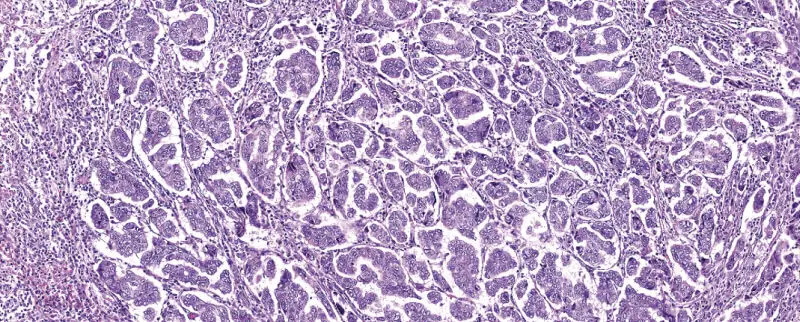
Invasive micropapillary carcinoma is a variant of ductal carcinoma characterized by an inside out morular growth pattern (Figure 1).1-3 The tumor cells consist of cell clusters that appear to float in a hollow space with associated retraction artifacts devoid of endothelial lining cells.2 The reversed polarity in these tumors can be demonstrated by an epithelial membrane antigen (EMA) stain, which shows positive cell membrane staining towards the stroma.1 The pseudopapillary clusters lack fibrovascular cores and comprise of cells that have intermediate to high grade nuclei, with variable pleomorphism containing macronucleoli and granular eosinophilic cytoplasm.2 The pure type, composed of 90% micropapillary features, accounts for 0.9-2.0% of all invasive carcinomas, whereas the mixed type, seen as a component of invasive carcinomas, amounts to 7.4%.1 Due to their association with lymphovascular tropism and lymph node metastases, these tumors exhibit aggressive behavior.1 The tumors may also be associated with apocrine and mucinous carcinomas of the breast and in the majority of cases, are ER and PR positive, while the positivity of HER2 can vary, ranging from 12.5% to 95%.1,4
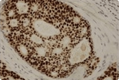
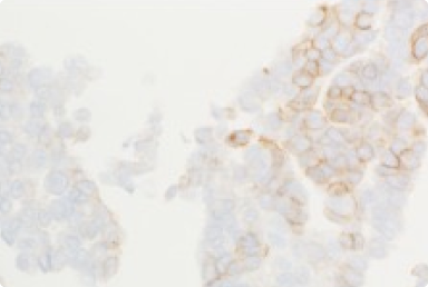
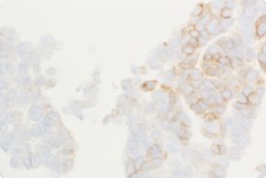
A basolateral HER2 IHC staining pattern is seen in invasive micropapillary carcinoma of the breast, corresponding to the basolateral lining of the cell membrane.1,2
“It is characterized by a linear C- or U-shaped phenomenon of HER2 staining that spares the luminal pole of the cell membrane (which faces the stroma due to the reversed polarity) (Figure 2).”1,2
The HER2 staining in these cases is incomplete and is not within the standard definitions to categorize HER2 expression, which define IHC 2+ staining as weak-to-moderate complete membrane staining observed in >10% of tumor cells with recommendation to perform reflex ISH testing.5 However, in addition to the standard definitions, the guidelines suggest that unusual staining patterns such as basolateral staining should be reported as IHC 2+ equivocal.5 In micropapillary carcinomas, the staining intensity can be variable between moderate and intense, and can be associated with HER2 gene amplification by FISH.6 In contrast, some invasive well-differentiated ductal carcinomas may exhibit weak or weak-to-moderate HER2 IHC basolateral staining but are only rarely associated with amplification.6 These cases usually occur due to the use of high sensitivity rabbit monoclonal anti-HER2 antibodies and should be interpreted with caution, to not overcall as equivocal (IHC 2+).6
The 2013 ASCO-CAP guidelines for HER2 testing stated that incomplete membranous staining that is faint or barely perceptible and in >10% of tumor cells was categorized as IHC 1+, while complete, weak-to-moderate membranous staining was categorized as IHC 2+.7 Invasive micropapillary carcinoma characterized by having an unusual basolateral HER2 immunohistochemical staining pattern shows incomplete moderate-to-strong staining and could not be compartmentalized into either of the 2013 IHC 1+ or IHC 2+ criteria.2
“This unusual IHC staining pattern led ASCO-CAP to modify their IHC 2+ recommendations in 2018 to weak-to-moderate complete membrane staining observed in >10% of tumor cells, and recommended follow up with reflex ISH testing.”5
The change was due to the fact that these carcinomas may harbor HER2 amplification.2 Furthermore, the guidelines also provide additional guidance for defining unusual staining patterns, like basolateral staining, such that if encountered, these cases should be reported as IHC 2+ equivocal.5 Basolateral staining may also rarely be seen in conventional invasive ductal carcinomas.6
When assessing HER2 IHC in invasive micropapillary carcinoma, it is important to pay attention to the staining pattern and intensity.1 These tumors predominantly demonstrate a basolateral staining pattern with an intensity that can be weak, moderate, or strong.1 In this setting, a weak-to-moderate HER2 stain should be reflexed to FISH.5 Cases with strong intensity usually correlate with HER2 gene amplification by FISH. In these cases, the HER2 intensity is more significant than the completeness of the HER2 IHC stain.2
ASCO, American Society of Clinical Oncology; CAP, College of American Pathologists; ER, estrogen receptor; EMA, epithelial membrane antigen; FISH, fluorescent in situ hybridization; HER2, human epidermal growth factor receptor 2; IHC, immunohistochemistry; IMPC, invasive micropapillary carcinoma; ISH, in situ hybridization; PR, progesterone receptor.
1. Perron M, et al. Arch Pathol Lab Med 2021; 145(8):979-987. 2. Zhou S, et al. Mod Pathol 2020; 33(7):1275-1286. 3. Yang YL, et al. Arch Pathol Lab Med 2016; 140(8):799-805. 4. Verras GI, et al. Breast Cancer (Dove Med Press) 2022; 14:41-61. 5. Wolff AC, et al. Arch Pathol Lab Med 2023;147(9):993-1000. 6. Bethune GC, et al. Histopathology 2017; 70(6):966-974. 7. Wolff AC, et al. J Clin Oncol 2018; 36(20):2105-2122.
Did you find this content useful?
Help us get it right next time by liking this article


©2025 Daiichi Sankyo, Inc. and AstraZeneca
US-84238, PP-US-8201a-1849; PP-US-8201a-2276, US-98135 03/25

Request an AstraZeneca Medical Affairs Representative Follow-Up
This website is intended to help healthcare professionals practicing in the United States find and access scientifically balanced, evidence-based, and peer-led information and professional resources in support of HER2 IHC evaluation. The dissemination of this information may be subject to different medical and regulatory requirements in other countries. This information is provided for educational purposes only. It is not intended for use in the diagnosis or treatment of individual patients. Are you a healthcare professional practicing in the United States?
I am a Daiichi Sankyo Company or AstraZeneca authorized person
This website is only intended for healthcare professionals practicing in the United States and authorized Daiichi Sankyo Company or AstraZeneca personnel. US Headquarters personnel and US Field Medical personnel are authorized to browse and search the database for their own background knowledge and insight. This website is not intended for use by US Field Sales personnel. None of the content of this site should be directly linked or distributed without approval.
