




Quick Links
Breast Cancer Links
- Breast Cancer Overview
- Breast Cancer Resources
- IHC Atlas
- Clinical Case Reviews
- Spotlight Series
- Knowledge Check


Welcome to the new, updated HER2Know
We have expanded our .
Looking for the original HER2Know breast cancer resources?


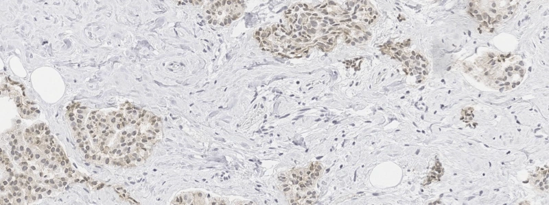
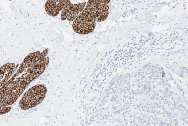
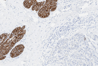
In this case, the HER2 IHC on a segmental mastectomy specimen from a primary invasive ductal carcinoma showed an unusual cytoplasmic staining pattern. HER2 was scored IHC 0. FISH was performed at an outside institution and was reported as negative ahead of the patient seeing a clinician. Without any pre-analytical or technical issues, the cause of the staining pattern is unclear, but it may include: HER2 internalization, cytoplasmic dimerization, phosphorylation, intensive HER2 transcription and presence of a cytoplasmic truncated HER2.1,2
As seen in Figure 1, the HER2 IHC shows diffuse cytoplasmic staining of mild intensity with no evidence of membranous staining. Based on these observations, the tumor would be categorized as IHC 0, as cytoplasmic HER2 staining should not be included in interpretation. FISH analysis of the tumor was negative for HER2 gene amplification (HER2/CEP17 ratio = 1.13, HER2 copy = 1.70).
The case demonstrates an unusual cytoplasmic HER2 staining pattern, which can make interpretation of this case challenging, as the presence of extensive cytoplasmic staining may hinder visualization and assessment of membrane staining of tumor cells.
The 2023 ASCO-CAP guideline update provides strict criteria for HER2 IHC categorization.3 A tumor can be scored as HER2 IHC 0 if there is no evidence of staining or incomplete, faint/barely perceptible membranous staining in <10% of tumor cells.3 Cytoplasmic staining is considered non-specific and should not be included in final HER2 IHC interpretation.4
Occasionally, diffuse cytoplasmic HER2 IHC staining can be encountered despite a lack of any pre-analytic issues. The cause of this artifactual staining may be unknown.1 In cases with cytoplasmic HER2 staining, subsequent ISH testing may be helpful.4
Some specific instances have been suggested to lead to the formation of this unusual cytoplasmic staining pattern. These include: HER2 internalization, cytoplasmic dimerization, phosphorylation, intensive HER2 transcription and presence of a cytoplasmic truncated HER2.1,2 In these instances, there is usually no evidence of HER2 gene amplification or increased levels of HER2 mRNA.1,2
Pathologists may find it helpful to familiarize themselves with cases that can be challenging to interpret. To explore more cases with challenging staining patterns, see pages 31 to 39 of HER2 expression in breast cancer: A comprehensive reference, found in the Breast Tumors Resources.
ASCO, American Society of Clinical Oncology; CAP, College of American Pathologists; CEP17, chromosome enumeration probe 17; ER, estrogen receptor; FISH, fluorescence in situ hybridization; HER2, human epidermal growth factor receptor 2; IHC, immunohistochemistry; ISH, in situ hybridization; Ki-67, proliferation marker; PR, progesterone receptor.
1. Blok EJ, et al. Clinical Medicine Insights: Oncology 2013;7:41-51. 2. Horiguchi S, et al. J Med Dent Sci 2010;57:155-163. 3. Wolff AC, et al. Arch Pathol Lab Med 2023;147(9):993-1000. 4. Wolff AC, et al. J Clin Oncol 2007;25(1):118-145.
Did you find this content useful?
Help us get it right next time by liking this article


©2025 Daiichi Sankyo, Inc. and AstraZeneca
PP-US-8201a-2248, PP-US-8201a-1849, PP-US-8201a-2036, US-84238, US-93987, US-102659 11/25

Request an AstraZeneca Medical Affairs Representative Follow-Up
This website is intended to help healthcare professionals practicing in the United States find and access scientifically balanced, evidence-based, and peer-led information and professional resources in support of HER2 IHC evaluation. The dissemination of this information may be subject to different medical and regulatory requirements in other countries. This information is provided for educational purposes only. It is not intended for use in the diagnosis or treatment of individual patients. Are you a healthcare professional practicing in the United States?
I am a Daiichi Sankyo Company or AstraZeneca authorized person
This website is only intended for healthcare professionals practicing in the United States and authorized Daiichi Sankyo Company or AstraZeneca personnel. US Headquarters personnel and US Field Medical personnel are authorized to browse and search the database for their own background knowledge and insight. This website is not intended for use by US Field Sales personnel. None of the content of this site should be directly linked or distributed without approval.
