





Quick Links
Breast Cancer Links
- Breast Cancer Overview
- Breast Cancer Resources
- IHC Atlas
- Clinical Case Reviews
- Spotlight Series
- Knowledge Check


Welcome to the new, updated HER2Know
We have expanded our .
Looking for the original HER2Know breast cancer resources?


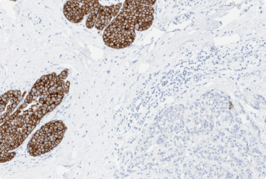
In this case, the HER2 IHC of a core needle biopsy specimen from an invasive breast carcinoma showed signs of edge artifact. Initial HER2 IHC was 2+, FISH negative. However, due to the presence of the artifact, repeat testing was performed. Repeat testing confirmed a HER2 IHC score of 2+ and negative FISH. This case will highlight the importance of avoiding HER2 IHC interpretation in areas with edge artifacts, and if extensive in amount to repeat testing if possible.* Core needle biopsy specimens in particular are prone to certain artifacts such as edge artifact and can make interpretation of these cases challenging.1
A core needle biopsy was performed on a 48-year-old female with a history of primary invasive breast carcinoma. HER2 IHC and FISH testing performed at an outside institution, revealed an IHC score of 2+ and negative FISH. However, it was observed that there was a significant edge artifact present on the sample.
The case was sent for consultation, and due to the presence of this significant edge artifact, HER2 IHC and FISH tests were repeated.
The term “edge artifact” is used to describe a staining pattern where there is focal strong staining present along the tissue edges or in regions where tissue is disrupted due to an unequal distribution of assay reagents.1 Edge artifacts can be caused by inconsistent fixation, the tissue drying out or lifting around the edges.2
Edge artifacts should be ignored during assay interpretation to avoid false-positive results.1 Tissue samples, particularly core needle biopsies, with significant edge artifact should be excluded from evaluation, and consideration should be given to repeating the test on a different sample.3
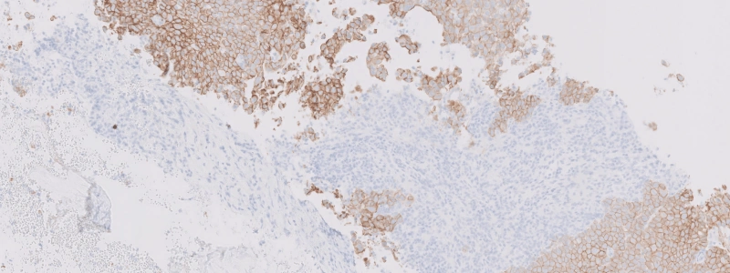
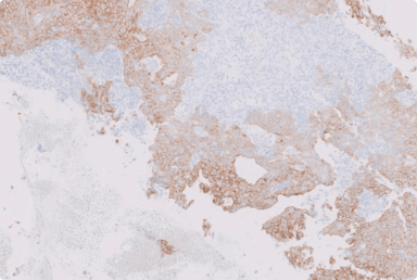
In the original HER2 IHC slide (Figure 1), most tumor cells show complete, circumferential membrane staining, ranging from moderate to strong intensity. However, there is a considerable edge artifact present in the core biopsy, with an appreciable variation in staining; where staining is highly concentrated at the peripheral edges of the tissue and diminishing in intensity towards the center of the tissue.
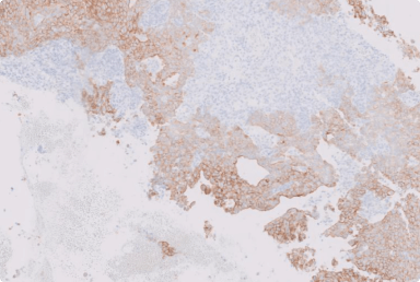
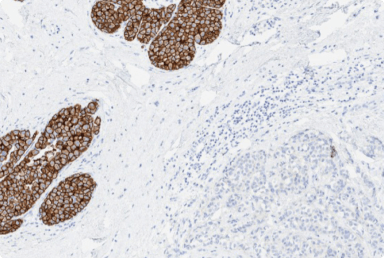
Repeat HER2 IHC testing (Figure 2) did not display the edge artifact shown previously, and instead shows a staining pattern that is consistent with HER2 IHC 2+, with most of the tumor cells showing complete, circumferential membranous staining with a weak-to-moderate intensity. Reflex HER2 FISH was negative.
It is important to be aware of artifacts that may hinder HER2 IHC interpretation, and can suggest problems with specimen processing and indicate poor quality staining.1,3 It is recommended to avoid interpreting HER2 IHC in cases with extensive edge artifacts.4 In such cases, repeat testing on a different block or specimen may be helpful.3 Certain sample types such as core needle biopsies are particularly prone to edge artifacts, so it is important to be mindful when evaluating these specimens.1
More guidance on how to approach similar challenging breast cancer cases can be found in the e-learning series titled Interpreting HER2 in breast cancer: A full spectrum of possibilities, found in the Breast Tumors Resources.
*These practice considerations represent the opinions of the Breast Pathology Faculty. Members of the Breast Pathology Faculty have been contracted as paid advisors through Daiichi Sankyo and AstraZeneca and have received compensation for their time.
CEP17, chromosome enumeration probe 17; ER, estrogen receptor; FISH, fluorescence in situ hybridization; HER2, human epidermal growth factor receptor 2; IHC, immunohistochemistry; PR, progesterone receptor.
1. Hicks DG and Schiffhauer L. Laboratory Medicine 2011;42(8):459-467. 2. VENTANA anti-HER2/neu (4B5) Rabbit Monoclonal Primary Antibody. Roche. 2022. 3. Wolff AC, et al. J Clin Oncol 2013;31(31):3997-4013. 4. Wolff AC, et al. J Clin Oncol. 2007;25(1):118-145.
Did you find this content useful?
Help us get it right next time by liking this article


©2025 Daiichi Sankyo, Inc. and AstraZeneca
PP-US-8201a-2248, PP-US-8201a-1849, PP-US-8201a-2036, US-84238, US-93987, US-102659 11/25

Request an AstraZeneca Medical Affairs Representative Follow-Up
This website is intended to help healthcare professionals practicing in the United States find and access scientifically balanced, evidence-based, and peer-led information and professional resources in support of HER2 IHC evaluation. The dissemination of this information may be subject to different medical and regulatory requirements in other countries. This information is provided for educational purposes only. It is not intended for use in the diagnosis or treatment of individual patients. Are you a healthcare professional practicing in the United States?
I am a Daiichi Sankyo Company or AstraZeneca authorized person
This website is only intended for healthcare professionals practicing in the United States and authorized Daiichi Sankyo Company or AstraZeneca personnel. US Headquarters personnel and US Field Medical personnel are authorized to browse and search the database for their own background knowledge and insight. This website is not intended for use by US Field Sales personnel. None of the content of this site should be directly linked or distributed without approval.
