



Quick Links
Breast Cancer Links
- Breast Cancer Overview
- Breast Cancer Resources
- IHC Atlas
- Clinical Case Reviews
- Spotlight Series
- Knowledge Check


Welcome to the new, updated HER2Know
We have expanded our .
Looking for the original HER2Know breast cancer resources?


This case is a mastectomy specimen of primary invasive lobular carcinoma, grade 2. There was an extensive in situ component present along with the invasive carcinoma, and HER2 IHC scores were discordant between the two. In situ components may be ductal or lobular in nature. Regardless, it is important to only evaluate the invasive component of the specimen and ignore all in situ components when determining a patient’s HER2 IHC score.1-3
A total mastectomy was performed on a 68-year-old female with primary invasive lobular carcinoma.
The cold ischemic time for the specimen was under 1 hour and fixation was in 10% NBF for 24 hours. Biomarkers were performed on the sample, and controls were all appropriate.
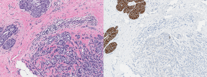
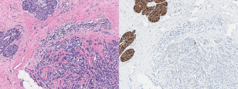
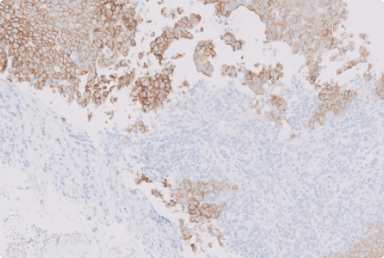
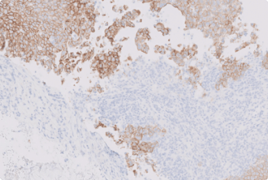
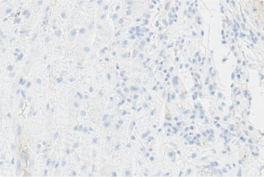
On the whole slide image (Figure 1), the area of invasive carcinoma shows no staining of HER2 (IHC 0). However, there is an extensive carcinoma in situ that shows complete and intense circumferential membrane staining. This can lead to misinterpretation of the specimen as HER2-positive, if not recognized as an in situ component.
The 2023 ASCO-CAP guideline update states that only the invasive component of a tumor should be evaluated for HER2 expression.1 In situ components should never be included in this evaluation.2 To aid in differentiating invasive and in situ components it is helpful to always review the H&E and IHC slides in conjunction.3
A visual guide with examples of whole slide images corresponding to each HER2 IHC score may help with HER2 assessment. HER2 differentiation in practice: a quick visual guide can be found in the Supporting Resources.
This case is challenging as there is discordance between HER2 IHC scores of the invasive carcinoma and the in situ component, with the in situ component potentially giving the appearance of a positive HER2 IHC result (false positive result). In situ components can be ductal or lobular in nature, DCIS or LCIS, respectively.4 It is important to be cognizant of the in situ components in invasive breast cancer IHC evaluation, as this component may be extensive in nature, and intermingled with the invasive component.2,3
It is important to always review the corresponding H&E slide together with the IHC slide and determine the invasive components of the specimen that should then be evaluated.3 In situ components can be apparent on H&E sections, however, in difficult cases IHC for myoepithelial markers can be used to distinguish the invasive and in situ components.5
Scroll through more Clinical Case Reviews for guidance on approaching challenging clinical cases.
*These practice considerations represent the opinions of the Breast Pathology Faculty. Members of the Breast Pathology Faculty have been contracted as paid advisors through Daiichi Sankyo and AstraZeneca and have received compensation for their time.
ASCO, American Society of Clinical Oncology; CAP, College of American Pathologists; DCIS, ductal carcinoma in situ; ER, estrogen receptor; H&E, hematoxylin and eosin stain; HER2, human epidermal growth factor receptor 2; IHC, immunohistochemistry; LCIS, lobular carcinoma in situ; NBF, neutral buffered formalin; PR, progesterone receptor.
1. Wolff AC, et al. Arch Pathol Lab Med 2023;147(9):993-1000. 2. Hicks DG and Schiffhauer L. Laboratory Medicine 2011;42(8):459-467. 3. Wolff AC, et al. J Clin Oncol 2007;25(1):118-145. 4. Makki J. Clinical Medicine Insights: Pathology 2015;8:23-31. 5. Zaha DC. World J Clin Oncol 2014;5(3):382-392.
Did you find this content useful?
Help us get it right next time by liking this article


©2025 Daiichi Sankyo, Inc. and AstraZeneca
PP-US-8201a-2248, PP-US-8201a-1849, PP-US-8201a-2036, US-84238, US-93987, US-102659 11/25

Request an AstraZeneca Medical Affairs Representative Follow-Up
This website is intended to help healthcare professionals practicing in the United States find and access scientifically balanced, evidence-based, and peer-led information and professional resources in support of HER2 IHC evaluation. The dissemination of this information may be subject to different medical and regulatory requirements in other countries. This information is provided for educational purposes only. It is not intended for use in the diagnosis or treatment of individual patients. Are you a healthcare professional practicing in the United States?
I am a Daiichi Sankyo Company or AstraZeneca authorized person
This website is only intended for healthcare professionals practicing in the United States and authorized Daiichi Sankyo Company or AstraZeneca personnel. US Headquarters personnel and US Field Medical personnel are authorized to browse and search the database for their own background knowledge and insight. This website is not intended for use by US Field Sales personnel. None of the content of this site should be directly linked or distributed without approval.
