

Quick Links
Breast Cancer Links
- Breast Cancer Overview
- Breast Cancer Resources
- IHC Atlas
- Clinical Case Reviews
- Spotlight Series
- Knowledge Check


Welcome to the new, updated HER2Know
We have expanded our .
Looking for the original HER2Know breast cancer resources?

In this case, the patient developed liver and bone metastases three years after initial diagnosis of Grade 1 invasive ductal carcinoma. A core liver biopsy from the metastatic sample was ER-negative (<1% nuclear staining), PR-positive (1% nuclear staining), with a score of IHC 0 with faint, incomplete staining (no ISH amplification). The primary tumor had a HER2 IHC 1+ score (also ISH negative), which highlights the importance of biomarker testing throughout the course of the disease as testing results are likely to vary between primary and metastatic sites.
A 56-year-old female was initially diagnosed with HR-positive, HER2-negative (IHC 1+), Grade 1 invasive ductal carcinoma. Three years after initial treatment with mastectomy and adjuvant hormone therapy, the cancer metastasized to liver and bone. At this stage, a core needle biopsy was performed on the metastatic site in the liver and the sample was processed for a repeat HER2 IHC testing.1
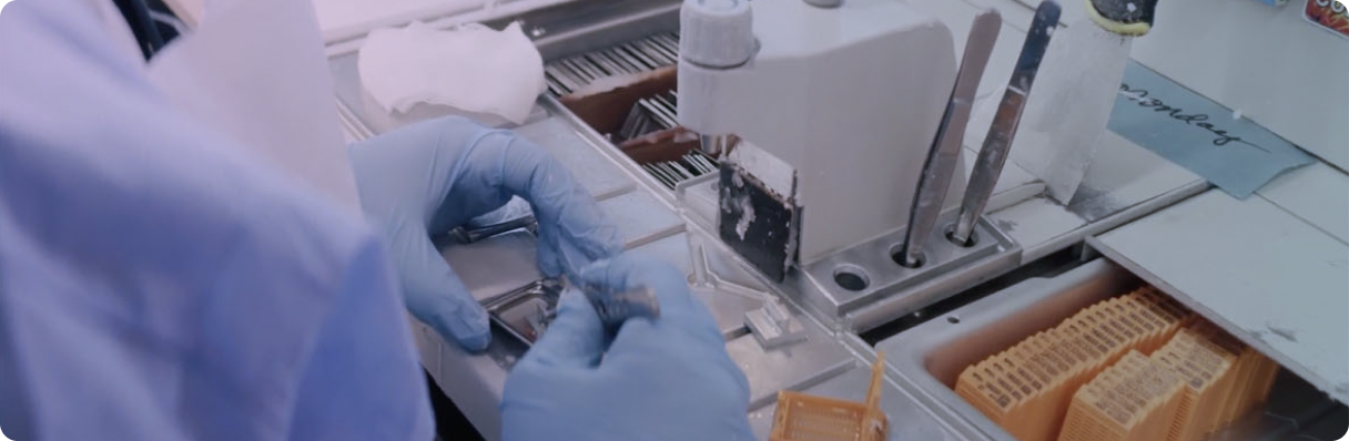
Pre-analytic variables may influence HER2 IHC results and should be considered in all specimen types from both the primary and metastatic sites.2 Deviations from standard pre-analytic variables may result in false-negative results.2-5 This is especially important when assessing HER2 IHC at the low end of the spectrum as a slight change in staining may impact treatment decisions.2,6
Pre-analytic variables should be carefully controlled, including:
Cold ischemic time and fixation
The following optimal tissue handling requirements are outlined in ASCO-CAP guidelines6:
Cold ischemic time should ideally be under 1 hour, as prolonged cold ischemic time may change HER2 IHC status.4,5,7 Prompt fixation preserves tissue quality and avoids compromising sample integrity due to rapid protein degradation after the interruption of the blood flow.2 Over- or under-fixation may affect the degree of protein cross-linking and antigen retrieval necessary for proper assay performance.2 This effect may also lead to incorrect hybridization in ISH testing and increase the chance of false-negative or false-positive results.2
Tissue processing
The following optimal tissue handling requirements are outlined in ASCO-CAP guidelines6:
Surgical resection specimens should be ideally sliced at 5-10 mm intervals to allow for optimal formalin penetration into the tissue; the chemical fixation should be uniform, with consistent fixative and fixation time.6
Optimally, HER2 staining should be performed on sections freshly cut from tissue blocks; however, if this is not feasible, the stored unstained sections should be stained within 6 weeks to ensure optimal preservation of antigenicity.6
In this case, a bone biopsy was not performed for HER2 IHC testing as the access to the metastatic site on the liver was more easily achieved. If processed appropriately, bone biopsy specimens are optimal for HER2 IHC testing: however, standardization of protocols and decalcification methods is necessary to avoid altering results.
Strong acids (e.g., hydrochloric acid) may rapidly macerate tissue and cause a loss of staining.8-10 Weak acids (e.g., formic acid) are slower in action and are more widely used for decalcification since they are less likely to interfere with immunostaining.8-10 Decalcification using chelating agents (e.g., EDTA) is best for preserving histomorphology and can be reliably used for IHC or ISH.8-10 However, they may be less suitable for urgent specimens due to longer processing time.9 Time for decalcification may also vary depending on the size or type of bone specimen (e.g., core biopsy, curettage, or resection).9
It is essential to validate the duration and the method of declassification, if decalcification of a bone specimen is deemed necessary, to ensure that they do not result in clinically relevant discordance.8 Prior to placing a specimen in the decalcification solution, consider removing any soft tissue within and/or adherent to bone and submit for histology processing without declassification. Often the soft tumoral tissues removed are sufficient for biomarker testing. Moreover, small amounts of bone spicules in the bone biopsies also can be processed normally without decalcification, and do not result in difficulty in the interpretation of histology or IHC.*
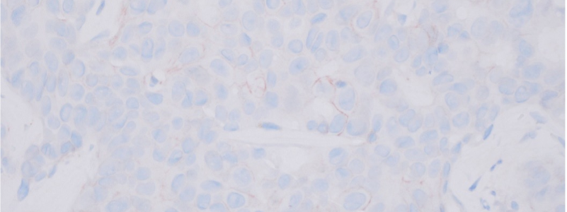
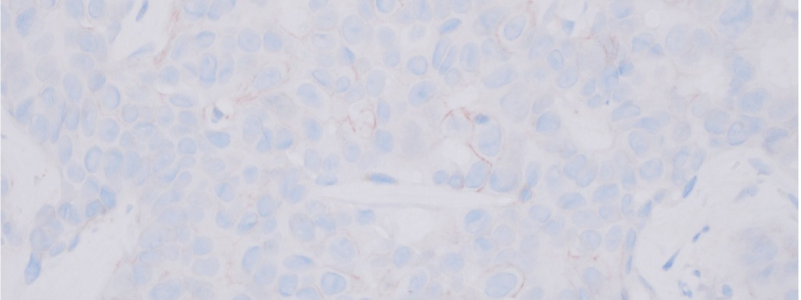
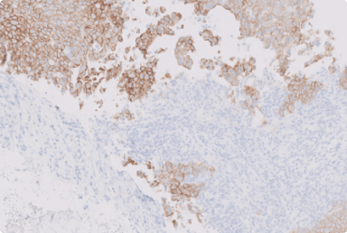
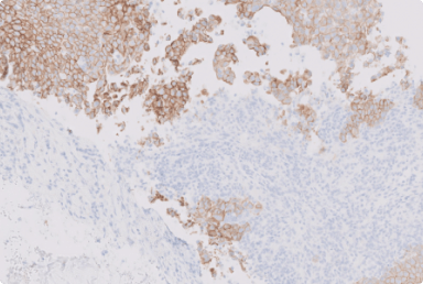
The HER2 IHC slide at 40X power of magnification shows faint/barely perceptible, incomplete membrane staining, which is found in around 5% of tumor cells.1 Based on the latest ASCO-CAP guideline update, this case is scored as IHC 0 with membrane staining that is incomplete and faint/barely perceptible in ≤10% of tumor cells.6
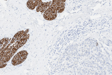
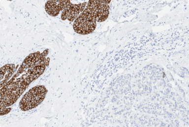
Estrogen receptor expression by immunohistochemistry:
Progesterone receptor expression by immunohistochemistry:
HER2 status by immunohistochemistry:
HER2 status by in situ hybridization (ISH):
Cold ischemia and fixation times
The specimen meets requirements specified in the latest version of the ASCO-CAP guidelines. The tissue is fixed at least for 6 hours but no more than 72 hours.
Technical notes
Formalin-fixed, paraffin embedded tissue is used. The tissue sections are stained with clone ER-SP1 for estrogen receptor, PR-IE2 for progesterone receptor, and 4B5 for HER2, using a biotin-free, polymer-based technology detection method and an automatic stainer.
Scoring for HER2 is based on the pattern and intensity of membranous staining. (Note: This assay has not been validated on decalcified tissues. If performed on a decalcified specimen, negative results should be interpreted with caution).
Accurate HER2 IHC interpretation is important to guide appropriate clinical decision making.6 Hence, it is key to carefully monitor pre-analytic variables to ensure they do not impact HER2 IHC results.
In this case, the HER2 IHC results changed from primary (IHC 1+) to metastatic breast cancer (IHC 0 with membrane staining that is incomplete and faint/barely perceptible in ≤10% of tumor cells).1 Change in HER2 status during the disease course is an observed event, occurring in about 10-30% of breast cancer cases.11-13 ASCO-CAP guidelines recommend considering HER2 IHC results on prior or concurrent primary samples (or other metastatic sites) as pre-analytic conditions may be better monitored in primary breast tissue samples than metastatic samples.6
You can find more information on how to approach the assessment of HER2 IHC 0 cases with faint, incomplete staining and IHC 0 without membrane staining in the Microscopy Series - Assessment of HER2 IHC in breast cancer: The lower end of HER2 expression, found here.
*These practice considerations represent the opinions of the Breast Pathology Faculty. Members of the Breast Pathology Faculty have been contracted as paid advisors through Daiichi Sankyo and AstraZeneca and have received compensation for their time.
ASCO, American Society of Clinical Oncology; CAP, College of American Pathologists; CEP17, chromosome 17 centromere; EDTA, ethylenediaminetetraacetic acid; ER, estrogen receptor; HER2, human epidermal growth factor receptor 2; HR, hormone receptor; IHC, immunohistochemistry; ISH, in situ hybridization; NOS, not otherwise specified; NST, no special type; PR, progesterone receptor.
1. Data on file. Daiichi Sankyo, Inc. Basking Ridge, NJ. 2. Tozbikian G, et al. Histopathology. 2024;85(3):489-502. 3. Khoury T, et al. Appl Immunohistochem Mol Morphol. 2014;22(9):688-695. 4. Yildiz-Aktas IZ, et al. Mod Pathol. 2012;25(8):1098-1105. 5. Khoury T, et al. Mod Pathol. 2009;22(11):1457-1467. 6. Wolff AC, et al. Arch Pathol Lab Med. 2023;147(9):993-1000. 7. Ivanova M, et al. Virchows Arch. 2024;484(1):3-14. 8. van Es SC, et al. Am J Surg Pathol. 2019;43(10):1355-1360. 9. Miquelestorena-Standley, et al. Mod Pathol. 2020;33:1505-1517. 10. Schrijver WA, et al. Mod Pathol. 2016;29(12):1460-1470. 11. Chen R, et al. Mod Pathol. 2020;33(12):2499-2506. 12. Schrijver WAME, et al. J Natl Cancer Inst. 2018;110(6):568-580. 13. Anderson S, et al. Breast Cancer Res Treat. 2023;201(3):437-446.
Did you find this content useful?
Help us get it right next time by liking this article


©2025 Daiichi Sankyo, Inc. and AstraZeneca
PP-US-8201a-2248, PP-US-8201a-1849, PP-US-8201a-2036, US-84238, US-93987, US-102659 11/25

Request an AstraZeneca Medical Affairs Representative Follow-Up
This website is intended to help healthcare professionals practicing in the United States find and access scientifically balanced, evidence-based, and peer-led information and professional resources in support of HER2 IHC evaluation. The dissemination of this information may be subject to different medical and regulatory requirements in other countries. This information is provided for educational purposes only. It is not intended for use in the diagnosis or treatment of individual patients. Are you a healthcare professional practicing in the United States?
I am a Daiichi Sankyo Company or AstraZeneca authorized person
This website is only intended for healthcare professionals practicing in the United States and authorized Daiichi Sankyo Company or AstraZeneca personnel. US Headquarters personnel and US Field Medical personnel are authorized to browse and search the database for their own background knowledge and insight. This website is not intended for use by US Field Sales personnel. None of the content of this site should be directly linked or distributed without approval.
