

HER2Know Logo
HER2Know Logo
  • Tumor Types
    • Welcome to the new, updated HER2Know

      We have expanded our resource library so you can now explore content on HER2 in tumors beyond breast.

      Looking for the original HER2Know breast cancer resources?

      • Breast Cancer
        • Breast Cancer Overview
        • Breast Cancer Resources
        • IHC Atlas
        • Pathologists' Perspectives
          • Clinical Case Reviews
          • Spotlight Series
        • Knowledge Check
      • Gastric Cancer
        • Gastric Cancer Overview
        • Gastric Cancer Resources
        • Gastric Cancer IHC Atlas
      • Other Solid Tumors
        • Other Solid Tumors Overview
        • Other Solid Tumors Resources
        • Other Solid Tumors IHC Atlas
       
  • Resources
  • IHC Atlas
  • Contributors
  • In the Know
  • Subscribe
  • TUMOR TYPES
  • Breast Cancer
    • Breast Cancer Overview
    • Breast Cancer Resources
    • IHC Atlas
    • Pathologists' perspectives
    • Clinical Case Reviews
    • Spotlight Series
    • Knowledge Check
  • Gastric Cancer
    • Gastric Cancer Overview
    • Gastric Cancer Resources
    • Gastric Cancer IHC Atlas
  • Other Solid Tumors
    • Other Solid Tumors Overview
    • Other Solid Tumors Resources
    • Other Solid Tumors IHC Atlas
  • Resources
  • IHC Atlas
  • Contributors
  • In the Know
  • Subscribe
  • ...
  • Pathologists' Perspectives
  • Clinical Case Reviews
  • Focal HER2 expression on IHC 0 breast carcinoma metastatic to the lung
  • Back to Clinical Case Reviews

Focal HER2 expression on IHC 0 breast carcinoma metastatic to the lung

Case contributed by Gary Tozbikian, MD, PhD

 
  •  LinkedIn
  • X
  • Facebook
The importance of reporting discrete HER2 IHC scores The importance of reporting discrete HER2 IHC scores
Breast Pathologist Dr Gary Tozbikian

Gary Tozbikian

MD

Case details

  • Age

    54
  • Gender

    Female

  • Primary/metastatic breast cancer

    Metastatic
  • Type of breast cancer

    Invasive ductal carcinoma NOS (NST)
  • Hormone receptor status

    ER: Positive (100% nuclear staining)
    PR: Positive (2% nuclear staining)
  • HER2 by IHC

    0 with faint, incomplete staining
  • Pre-analytic variables and technical issues

    None
  • Specimen type in metastatic setting

    Lung biopsy
  • Prior test results

    Reported history of primary breast cancer diagnosed 10 years prior. The primary breast cancer was a Grade 1, invasive ductal
    carcinoma, ER: 50%, PR: 70%, HER2 IHC 0
    (HER2-negative)

In this case, a patient presented with invasive ductal carcinoma that metastasized to the lung. HER2 IHC on the tumor biopsy showed HER2 expression that can be described as IHC 0 with faint, incomplete staining.1 Examining the entire slide at high power is needed to detect focal staining on tumors with low levels of HER2 expression.1,2*

Case summary

A 54-year-old female was diagnosed with HR-positive invasive ductal carcinoma that had progressed to metastatic disease.1 The patient was previously diagnosed with Grade 1, HR-positive, HER2-negative primary breast cancer 10 years prior. At metastatic diagnosis, lung biopsy was obtained and analyzed for key biomarkers.1

How were pre-analytic and analytic variables controlled for the case?

The lung biopsy sample was immediately placed into neutral buffered formalin and fixed for 12 hours and 49 minutes.1 HER2 IHC testing was performed with Ventana PATHWAY anti-HER2/neu 4B5 assay.1

What does the HER2 IHC staining indicate?

Before revealing the answer, attempt interpretation of the slide by viewing the whole slide image here.

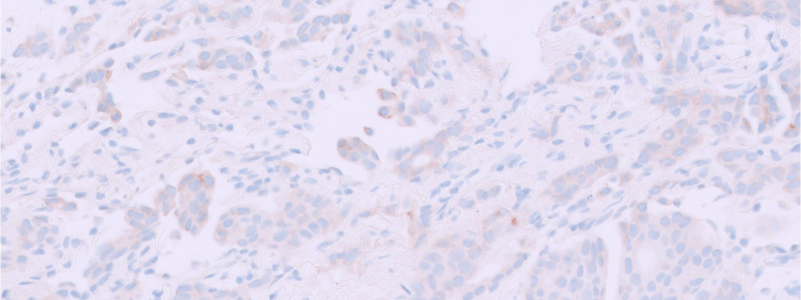
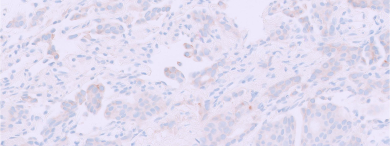
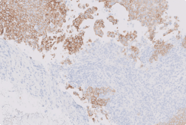
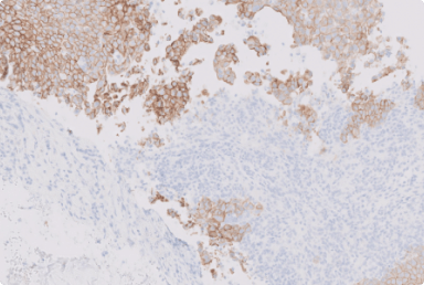
The slide shows intratumoral heterogeneity for HER2 expression.1 We can see focal staining at 40X power of magnification that shows faint/barely perceptible, incomplete membrane staining that is in less than 10% of tumor cells.1

Figure 1. Focal faint/barely perceptible, incomplete HER2 IHC membrane staining (40X)1

Click here to view and interact with whole slide images

As recommended by ASCO-CAP guidelines, a second pathologist reviewed the case to confirm the accuracy of the score at the lower end of the spectrum. An emphasis was made that faint cytoplasmic staining, present in this case, should not be considered in the assessment.2,3

How were the results reported?*

Estrogen receptor:

Positive (100%, strong intensity)

Progesterone receptor:

Positive (2%, moderate intensity)

HER2:

Negative (Score 0 / Membrane staining that is incomplete and is faint/barely perceptible and in less than or equal to 10% of tumor cells (0+/with membrane staining))

Comment:

Per the electronic medical record, the patient's history of breast cancer and colorectal cancer is noted. Immunohistochemical stains show that the tumor is positive for CK7 and GATA3, and is negative for CK20, TTF-1, CDX2, and SATB2. The findings are consistent with a metastasis of breast origin.

“Detecting HER2 IHC 0 with faint, incomplete staining requires careful interrogation of the entire tumor at high power (40X) to identify focal areas of HER2 reactivity.”

Discussion

The current case was reported as HER2-negative (IHC 0 with faint, incomplete staining), which is consistent with the primary breast cancer.1 While in the metastatic setting there was evidence of focal membrane staining, it is unclear, from the report, if the previous tumor showed any level of HER2 staining.1

IHC 0 can be defined as membrane staining that is incomplete and is faint/barely perceptible and in ≤10% of tumor cells (0+) or no membrane staining (0). Staining on IHC 0 tumors can often be focal, which can make the assessment challenging.1 Hence, scanning the entire tumor at high power (40X) is needed to detect focal HER2 reactivity and distinguish IHC 0 with faint, incomplete staining from IHC 0 without membrane staining.1,2*

Considerations

  • Assess HER2 IHC at 40X power of magnification to detect faint and incomplete staining patterns2
  • Scan the entire tumor to detect focal membrane staining1*
  • Discount the presence of cytoplasmic (blush) staining when evaluating and scoring HER2 expression by IHC2,3
  • Report intensity, pattern and percentage of the tumor cell staining as it may inform clinical decision making4

You can find more information on how to approach assessment of HER2 IHC 0 with faint, incomplete staining in the Microscopy Series - Assessment of HER2 IHC in breast cancer: The lower end of HER2 expression, by clicking here.

*These practice considerations represent the opinions of the Breast Pathology Faculty. Members of the Breast Pathology Faculty have been contracted as paid advisors through Daiichi Sankyo and AstraZeneca and have received compensation for their time.


ASCO, American Society of Clinical Oncology; CAP, College of American Pathologists; CDX2, caudal type homeobox 2; CK7, cytokeratin 7; CK20, cytokeratin 20; ER, estrogen receptor; GATA3; GATA binding protein 3; HER2, human epidermal growth factor receptor 2; HR, hormone receptor; IHC, immunohistochemistry; NOS, not otherwise specified; NST, no special type; PR, progesterone receptor; TTF-1; thyroid transcription factor -1.


1. Data on file. Daiichi Sankyo, Inc. Basking Ridge, NJ. 2. Wolff AC, et al. Arch Pathol Lab Med. 2023;147(9):993-1000. 3. Grassini D, et al. Pathobiology. 2022;89:278-296. 4. Ivanova M, et al. Virchows Arch. 2024;484(1):3-14.

Did you find this content useful?
Help us get it right next time by liking this article

.

Explore more Clinical Case Reviews

View all

In this series of articles, the Pathology Faculty share their experience with complex clinical cases and highlight practices that help to aid consistency in HER2 IHC scoring.

View all
assessing-her2-ihc-with-cytoplasmic-staining.png

Assessing HER2 IHC with cytoplasmic staining

In this case, the HER2 IHC on a segmental mastectomy specimen from a primary invasive ductal carcinoma showed an unusual cytoplasmic staining pattern.

by Savitri Krishnamurthy, MD

 
  •  LinkedIn
  • X
  • Facebook
assessing-her2-ihc-with-edge-artifacts.png

Assessing HER2 IHC with edge artifacts

In this case, the HER2 IHC of a core needle biopsy specimen from an invasive breast carcinoma showed signs of edge artifact.

by Gary Tozbikian, MD

 
  •  LinkedIn
  • X
  • Facebook
interpreting-her2-ihc-with-an-extensive-in-situ-component.png

Interpreting HER2 IHC with an extensive in situ component

In this case, there was an extensive in situ component present along with the invasive carcinoma, and HER2 IHC scores were discordant between the two.

by Hannah Wen, MD, PhD

 
  •  LinkedIn
  • X
  • Facebook
Her2know Logo

Quick Links

  • Subscribe
  • Resources
  • Contributors


Breast Cancer Links

  • Breast Cancer Overview
  • Breast Cancer Resources
  • IHC Atlas
  • Clinical Case Reviews
  • Spotlight Series
  • Knowledge Check


Gastric Cancer Links

  • Gastric Cancer Overview
  • Gastric Cancer Resources

About Us

  • Contact Us
  • Request Medical Follow-Up
  • AstraZeneca
  • AZ Medical
  • Daiichi Sankyo Company
  • DSI Medical
  • Your Privacy Choices

Legal

  • Privacy Notice
  • Cookie Preferences
  • Legal Notices & Terms of Use
  • Consumer Health Data Privacy Notice
.
Daiichi-Sankyo - AstraZeneca Footer

©2025 Daiichi Sankyo, Inc. and AstraZeneca
PP-US-8201a-2248, PP-US-8201a-1849, PP-US-8201a-2036, US-84238, US-93987, US-102659 11/25

HER2Know Logo

Request an AstraZeneca Medical Affairs Representative Follow-Up

Welcome to HER2Know

This website is intended to help healthcare professionals practicing in the United States find and access scientifically balanced, evidence-based, and peer-led information and professional resources in support of HER2 IHC evaluation. The dissemination of this information may be subject to different medical and regulatory requirements in other countries. This information is provided for educational purposes only. It is not intended for use in the diagnosis or treatment of individual patients. Are you a healthcare professional practicing in the United States?

I am a Daiichi Sankyo Company or AstraZeneca authorized person

What is a Daiichi Sankyo Company or AstraZeneca authorized person?

This website is only intended for healthcare professionals practicing in the United States and authorized Daiichi Sankyo Company or AstraZeneca personnel. US Headquarters personnel and US Field Medical personnel are authorized to browse and search the database for their own background knowledge and insight. This website is not intended for use by US Field Sales personnel. None of the content of this site should be directly linked or distributed without approval.

Self Certification Buttons
No, take me to AstraZeneca.com Self Certification Buttons